PET imaging of hypoxia in tumours
Most solid tumours have a varying amount of hypoxic cells, representing viable cells adapted to a low oxygen concentration. Near-zero oxygen concentrations are often found adjacent to regions of tumour necrosis. The cells in a hypoxic microenvironment are resistant to radiotherapy and less accessible to several chemotherapeutic drugs. Hypoxic environment selects for invasive, more malignant phenotypes. Hypoxia promotes angiogenesis in tumours as well as in healthy tissue. Acute hypoxia occurs when the microvessels surrounding the tissue close up, usually transiently. Chronic hypoxia occurs frequently in growing tumours when part of the tumour cells are far from functional capillaries, or uncontrolled cell proliferation increases the demand of oxygen. Tumour hypoxia is thus spatially and temporally heterogeneous.
Measuring the hypoxic fraction in tumours enhances the development of hypoxia-targeted therapy (Brown, 2007; Minn et al., 2008). Measurement of hypoxia may also be useful in patients with acute ischemic stroke (Read et al., 1998; Alawneh et al., 2014; Lee et al., 2014). Renal hypoxia is implicated in the development of chronic kidney disease (Ow et al., 2018), and adipose tissue hypoxia in the metabolic syndrome.
Tissue hypoxia can be assessed by pO2 probes, but PET measurement is non-invasive and potentially provides 3D image of chronically hypoxic tissue region (Markus et al., 2002). A number of PET radioligands have been developed and validated, most of them 2-nitroimidazole derivatives. [18F]FMISO, [64Cu]ATSM, and many other hypoxia PET radiopharmaceuticals are significantly taken up only in cells in extreme hypoxic environments at oxygen levels that are well below levels that induce radioresistance and HIF1α expression (Chia et al., 2022). This may hamper their use in radiotherapy planning and clinical trials of hypoxia modification strategies.
Li et al. (2014) reviewed the modelling methods for the most commonly used PET tracers for hypoxia.
Nitroimidazoles
2-Nitroimidazoles are the most widely studied compounds for imaging hypoxia (Nunn et al., 1995). Nitroimidazoles are reduced intracellularly in all viable cells and have an established use in the treatment of anaerobic infections. In aerobic cells the reduced nitroimidazole is immediately re-oxidised and washed out rapidly. By contrast, in cells with a low oxygen concentration the re-oxidation is slowed, which allows further reductive reactions to take place. This leads to the formation of reactive products that can covalently bind to cell components or are charged and thus diffuse more slowly out of the tissue (Nunn et al., 1995). Thus, hypoxia increases both reversible and irreversible components of nitroimidazole uptake in tissue.
2-Nitroimidazole derived PET radiopharmaceuticals include [18F]FMISO (Casciari et al., 1995; Thorwarth et al., 2005; Wang et al., 2009), [18F]FETNIM (Lehtiö et al., 2003, 2004; Grönroos et al., 2014), [18F]EF5 (Komar et al., 2008; Silvoniemi et al., 2014; Silén et al., 2014; Silvoniemi et al., 2017), [18F]FAZA (Verwer et al., 2014), and [18F]HX4 (also called [18F]flortanidazole) (Verwer et al., 2017).
Reduction rate of 2-nitroimidazoles is dependent on both oxygen partial pressure and levels of nitroreductase enzyme levels, so that for example [18F]FMISO imaging may be detecting not only severely hypoxic tumours but also mildly hypoxic tumours with high nitroreductase activity (Koch & Evans, 2015).
Casciari et al. (1995) have developed a kinetic compartmental model to quantify tumour oxygenation status from PET data using fluorine-18 labelled fluoromisonidazole ([18F]FMISO), which is the most widely used nitroimidazole compound in clinical PET. Thereafter, [18F]FMISO model has been reduced to irreversible two-tissue compartment model (Thorwarth et al., 2005; Wang et al., 2009; Grkovski et al., 2017; McGowan et al., 2017). Reversible two-tissue compartmental model was found to provide best fits for [18F]HX4 (Verwer et al., 2017).
In clinical applications, late-scan protocols are usually followed, without blood sampling. Data is analysed using robust semi-quantitative methods such as SUV and tumour-to-muscle ratio (T/M). [18F]EF5 T/M has excellent repeatability in head and neck cancer (Silvoniemi et al., 2018). Tumour-to-blood ratio would be better than tumour-to-muscle ratio, because of considerable variability in muscle uptake. When heart LV cavity is not in the field-of-view, muscle could be used as surrogate for the blood by accounting for the fat content of the muscle by incorporating CT HU in the SUV ratio equation (Han et al., 2018).
Redox state tracers
Intracellular redox environment is altered as a consequence of hypoxia, as the concentration of reducing agents such as NADH are increased. Several metal chelates that are sensitive to the redox state have been developed and labelled, usually based on the reduction of Cu2+ to Cu+. For example, [64Cu]ATSM is highly membrane permeable and has rapid washout from non-hypoxic regions, but it is retained in hypoxic tissue (Lewis et al., 1999). However, retention of [64Cu]ATSM and also [64Cu]PTSM was shown to be reduced in tumours that express MDR1 protein (Liu et al., 2009), limiting the clinical applications of these radiopharmaceuticals.
Carbonic anhydrase IX
Carbonic anhydrases catalyze the reversible hydration of CO2 to HCO3- and H+ or H2CO3, helping in regulation of pH and CO2 transport. Expression of carbonic anhydrase IX (CA-IX) is upregulated in hypoxia through hypoxia inducible factor-1 (HIF-1) cascade. CA-IX is involved in cancer development, and is therefore considered as target in clinical imaging and radiotherapy.
[18F]FDG
[18F]FDG is widely used PET tracer for cancer studies because of the increased demand of glycolysis in most tumours. Increased [18F]FDG uptake is often seen in hypoxic tissues and it may correlate well with the expression of hypoxia-inducible factors. However [18F]FDG uptake is not specific to hypoxia, but a result of many factors such as inflammation and angiogenesis (Dierckx and Van de Wiele, 2008).
Combination of perfusion and [18F]FDG PET studies can be used to quantify perfusion-metabolism mismatch.
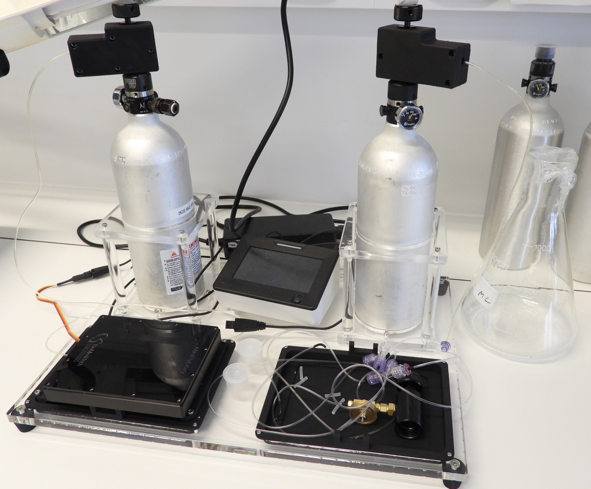
Carbogen breathing increases tumour oxygenation, and it was shown to reduce [18F]FDG uptake in a mouse model (Neveu et al., 2015).
MCT4
HIF-1-α controls the expression of monocarboxylase transporter MCT4, which allows export of lactate by glycolytic and hypoxic cells and maintenance of intracellular pH homeostasis (Gündel et al., 2021). Cytosolic carbonic anhydrase II enhances transport activity of MCT1 and MCT4, while extracellular membrane-bound carbonic anhydrase IV increases transport activity of MCT2 (Adeva-Andany et al., 2014).
See also:
- [18F]EF5
- Perfusion-metabolism mismatch volume
- Mitochondria
- Angiogenesis
- Erythropoietin (EPO)
- Apoptosis
- Reactive oxygen species (ROS)
Literature
Carreau A, El Hafny-Rahbi B, Matejuk A, Grillon C, Kieda C. Why is the partial oxygen pressure of human tissues a crucial parameter? Small molecules and hypoxia. J Cell Mol Med. 2011; 15(6): 1239-1253. doi: 10.1111/j.1582-4934.2011.01258.x.
Dhani N, Fyles A, Hedley D, Milosevic M. The clinical significance of hypoxia in human cancers. Semin Nucl Med. 2015; 45: 110-121. doi: 10.1053/j.semnuclmed.2014.11.002.
Fleming IN, Manavaki R, Blower PJ, West C, Williams KJ, Harris AL, Domarkas J, Lord S, Baldry C, Gilbert FJ. Imaging tumour hypoxia with positron emission tomography. Br J Cancer 2015; 112: 238-250. doi: 10.1038/bjc.2014.610.
Frezza C, Gottlieb E. Mitochondria in cancer: not just innocent bystanders. Semin Cancer Biol. 2009; 19: 4-11. doi: 10.1016/j.semcancer.2008.11.008.
Höckel M, Vaupel P. Tumor hypoxia: definitions and current clinical, biologic, and molecular aspects. J Natl Cancer Inst. 2001; 93: 266-276. doi: 10.1093/jnci/93.4.266.
Kelada OJ, Carlson DJ. Molecular imaging of tumor hypoxia with positron emission tomography. Radiat Res. 2014; 181: 335-349. doi: 10.1667/RR13590.1.
Krohn KA, Link JM, Mason RP. Molecular imaging of hypoxia. J Nucl Med. 2008; 49(6): 129S-148S. doi: 10.2967/jnumed.107.045914.
Li F, Joergensen JT, Hansen AE, Kjaer A. Kinetic modeling in PET imaging of hypoxia. Am J Nucl Med Mol Imaging 2014; 4(5): 490-506. PMID: 25250200.
Shi K, Bayer C, Astner ST, Gaertner FC, Vaupel P, Schwaiger M, Huang S-C, Ziegler SI. Quantitative analysis of [18F]FMISO PET for tumor hypoxia: correlation of modeling results with immunohistochemistry. Mol Imaging Biol. 2017; 19(1): 120-129. doi: 10.1007/s11307-016-0975-4.
Span PN, Bussink J. Biology of hypoxia. Semin Nucl Med. 2015; 45: 101-109. doi: 10.1053/j.semnuclmed.2014.10.002.
Taylor E, Gottwald J, Yeung I, Keller H, Milosevic M, Dhani NC, Siddiqui I, Hedley DW, Jaffray DA. Impact of tissue transport on PET hypoxia quantification in pancreatic tumours. EJNMMI Res. 2017; 7(1): 101. doi: 10.1186/s13550-017-0347-3.
Verwer EE, Boellaard R, van der Veldt AAM. Positron emission tomography to assess hypoxia and perfusion in lung cancer. World J Clin Oncol. 2014; 5(5): 824-844. doi: 10.5306/wjco.v5.i5.824.
Verwer EE, Zegers CM, van Elmpt W, Wierts R, Windhorst AD, Mottaghy FM, Lambin P, Boellaard R. Pharmacokinetic modeling of a novel hypoxia PET tracer [18F]HX4 in patients with non-small cell lung cancer. EJNMMI Phys. 2016; 3(1): 30. doi: 10.1186/s40658-016-0167-y.

Tags: Hypoxia, Perfusion, Oxygen, Tumour, Angiogenesis, Carbonic anhydrase
Updated at: 2023-08-01
Created at: 2007-10-23
Written by: Vesa Oikonen