Image reconstruction
Reconstruction is the process where actual PET image is formed from the raw data (sinograms) collected by the PET scanner.
PET images are normally reconstructed from the raw data by the personnel of PET centre using the software provided by scanner manufacturers, and images are stored in PACS.
Filtered-back-projection (FBP) reconstruction is based on the classic analytic algorithm, and is fast and robust, but cannot account for the advanced data acquisition methods of the current PET scanners. Modern iterative algorithms such as maximum-likelihood expectation maximization (ML-EM) and a faster version of it, ordered subset expectation maximization (OSEM) may improve the quality of the reconstructed image, but are slower to compute, and reconstruction parameters must be carefully selected and verified. Inappropriate selection of reconstruction parameters will affect the quantitative image values (Jaskowiak et al., 2005; Vriens et al., 2010; Matheoud et al., 2011). If input function is derived from the dynamic PET image, then that must be considered in the reconstruction; OSEM can reduce noise, but also cause bias in the image-based input function, when compared to FBP (Feng et al., 2015).
Image reconstruction involves numerous forward and backward projection calculations, which is computationally intensive process, but can be easily parallelized to computing clusters or GPUs (Després et al., 2017).
Reconstruction of ECAT 931 images
The sinograms of the old ECAT 931 studies are stored by the PET Centre, but the storing of the PET images were on the responsibility of the research group. Researchers are strongly recommended to search for the PET images from their old storage and backup disks, instead of trying to reconstruct the data again.
Reconstruction would now be a laborious and difficult process, because the original workstations with original software are no more available.
Process
Previously, GUI program for reconstruction of ECAT 931 and 2D GE Advance sinograms, both stored in CTI ECAT 6.3 format, was found in the menus on SUN/Solaris platforms. This is no longer functional, but command-line programs for normalization and attenuation correction, FBP and MRP reconstruction, decay correction, and image calibration are available for Windows, Linux, and macOS. Older command-line versions of reconstruction and calibration correction programs can be used from SUN/Solaris Unix command-prompt, if an old Solaris workstation is still available.
Required data
For reconstruction you need obviously the sinograms (*.scn), but
you also need the attenuation and normalization files (*.atn and
*.nrm). Files may be found in folder
S:\Archive\Scanners\Ecat\ecat-datnauhat\.
After reconstruction, images need to be calibrated to units kBq/mL, and for that you need appropriate calibration file (see below).
Reconstruction in Windows, Linux, or macOS
Open the command prompt window in your system, or write a script for doing the necessary steps, normalization and attenuation correction, FBP or MRP reconstruction, decay correction, and image calibration. For example:
ecatnorm s04065dy2.scn 991005_norm.nrm s04065tr2.atn s04065dy2_corrected.scn
ecatfbp -dim=192 -zoom=1.5 s04065dy2_corrected.scn s04065dy2.img
imgdecay -dd s04065dy2.img
In this example, the data is from a late-scan (dy2) and the decay correction would still need to be corrected to the time of radioligand administration.
This example used FBP reconstruction, but procedure for MRP reconstruction would be similar except of the program name and some optional parameters.
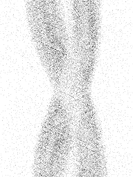

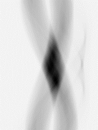
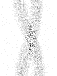
Figure 1a. Example of ECAT 931 data from a leg study: emission sinogram, normalization sinogram, attenuation correction data, and normalization and attenuation corrected emission sinogram. Single frame and plane #7.
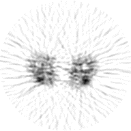

Figure 1b. Examples of images reconstructed from the normalization and attenuation corrected emission sinogram (Figure 1a) using FBP and MRP.
Reconstrion on SUN/Solaris
FBP reconstruction:
Open remote login window to Jupiter (or whatever available Solaris workstation). The FBP reconstruction program fbprec is called from command prompt window with command
/pet-storage/petsoftware/bin/unix/old/fbprec parameter_file.txt
The format of fbprec parameter file is specified here.
MRP reconstruction:
Open remote login window to the Solaris workstation. The MRP reconstruction (Alenius and Ruotsalainen, 1997, 1998, 2002) program mrprec is called from command prompt window with command
/pet-storage/petsoftware/bin/unix/old/mrprec parameter_file.txt
The format of mrprec parameter file is specified here.
Calibration
Reconstructed image still needs to be calibrated to units kBq/mL with ecalibr; this is done with command
ecalibr imagefile calibrationfile
or
ecalibr imagefile calibrationfilepath
Calibration files are currently stored in
S:\lab\plasma\ECAT931\plane_calibration.dir
and they are named as YYMMDD; Calibrations done before 1997 are located in folder
.old with names such as 13-APR-1993.
Calibration files (example) contain the calibration measurement
date, calibration coefficients for the 15 image planes, and the mean of calibration coefficients,
each on their own line.
You must select the calibration that was performed on the PET scan day, or the previous calibration,
or if PET scan is from year 1997 or later, let the program select the file from the path based on
scan start date found in the image header and the name of the calibration file.
Scan date can be determined from patient records or from sinogram file with egetstrt.
Continuing the example study above, the calibration command would be:
ecalibr s04065dy2.img S:\Lab\plasma\ECAT931\plane_calibration.dir\991005
Reconstruction of GE Advance 2D images
Normally, GE Advance images are reconstructed on the GE Advance workstation. However, 2D sinograms can be extracted in ECAT 6.3 sinogram format, and these can be reconstructed equally. However, separate attenuation and normalization files are not needed, because these corrections are included in GE Advance sinograms.
Resulting image is not calibrated automatically. Calibration can be done using program calib4ge. Windows program for calibration does not yet exist.
Re-projection of images
For testing purposes, it is possible to compute a re-projected sinogram using program img2scn.
References
Alenius S, Ruotsalainen U. Bayesian image reconstruction for emission tomography based on median root prior. Eur J Nucl Med. 1997; 24: 258-265. doi: 10.1007/BF01728761.
Alenius S, Ruotsalainen U, Astola J. Using Local Median as the Location of the Prior Distribution in Iterative Emission Tomography Image Reconstruction. IEEE Trans Nucl Sci. 1998; 45(6): 3097-3104. doi: 10.1109/23.737670.
Alenius S, Ruotsalainen U. Generalization of Median Root Prior Reconstruction. IEEE Trans Med Imaging 2002; 21(11): 1413-1420. doi: 10.1109/TMI.2002.806415.
Bettinardi V, Alenius S, Numminen P, Teras M, Gilardi MC, Fazio F, Ruotsalainen U. Implementation and evaluation of an ordered subsets reconstruction algorithm for transmission PET studies using median root prior and inter-update median filtering. Eur J Nucl Med. 2003; 30(2): 222-231. doi: 10.1007/s00259-002-1046-4.
Bettinardi V, Pagani E, Gilardi MC, Alenius S, Thielemans K, Teras M, Fazio F. Implementation and evaluation of a 3D one-step late reconstruction algorithm for positron emission tomography brain studies using median root prior. Eur J Nucl Med. 2002; 29(1): 7-18. doi: 10.1007/s002590100651.
Dahlbom M (ed.): Physics of PET and SPECT Imaging. CRC Press, 2017. ISBN 978-1-4665-6013-0.
Després P, Jia X. A review of GPU-based medical image reconstruction. Phys Med. 2017; 42: 76-92. doi: 10.1016/j.ejmp.2017.07.024.
Herman GT: Fundamentals of Computerized Tomography. Image Reconstruction from Projections. Springer, 2009. doi: 10.1007/978-1-84628-723-7.
Jan J: Medical Image Processing, Reconstruction and Restoration - Concepts and Methods. CRC Press, 2006, ISBN: 978-0-8247-5849-3.
Johansson J. Tomografiamenetelmän käänteisongelman ratkaisusta. Pro gradu, 2007.
Johansson J. Radon Transform in Positron Emission Tomography.
Kinahan PE, Defrise M, Clackdoyle R (2004): Analytic image reconstruction methods. In: Emission Tomography: The Fundamentals of PET and SPECT. (Eds: Wermick MN, Aarsvold JN). Elsevier Inc., pp 421-442.
Lalush DS, Wernick MN (2004): Iterative image reconstruction. In: Emission Tomography: The Fundamentals of PET and SPECT. (Eds: Wermick MN, Aarsvold JN). Elsevier Inc., pp 443-472.
Lindén J. Positroniemissiotomografian rekonstruktioalgoritmeista ja niiden simulaatiotutkimuksista. Pro gradu, 2013.
Markiewicz PJ, Ehrhardt MJ, Erlandsson K, Noonan PJ, Barnes A, Schott JM, Atkinson D, Arridge SR, Hutton BF, Ourselin S. NiftyPET: a high-throughput software platform for high quantitative accuracy and precision PET imaging and analysis. Neuroinformatics 2018; 16(1): 95-115. doi: 10.1007/s12021-017-9352-y.
Rahmim A, Tang J, Zaidi H. Four-dimensional (4D) image reconstruction strategies in dynamic PET: beyond conventional independent frame reconstruction. Med Phys. 2009; 36(8): 3654-3670. doi: 10.1118/1.3160108.
STIR: Software for Tomographic Image Reconstruction. homepage.
Teräs M, Tolvanen T, Johansson JJ, Williams JJ, Knuuti J. Performance of the new generation of whole-body PET/CT scanners: Discovery STE and Discovery VCT. Eur J Nucl Med Mol Imaging 2007; 34: 1683-1692. doi: 10.1007/s00259-007-0493-3.
Walker MD, Asselin M-C, Julyan PJ, Feldmann M, Talbot PS, Jones T, Matthews JC. Bias in iterative reconstruction of low-statistics PET data: benefits of a resolution model. Phys Med Biol. 2011; 56: 931-949. doi: 10.1088/0031-9155/56/4/004.
Wettenhovi V-V, Vauhkonen M, Kolehmainen V. OMEGA - Open-source emission tomography software. Phys Med Biol. 2021; 66(6): 065010. doi: 10.1088/1361-6560/abe65f.
Tags: Sinogram, Image reconstruction, Calibration, Attenuation, Normalization, ECAT, PET, Image
Updated at: 2021-02-18
Created at: 2009-02-20
Written by: Vesa Oikonen, Kaisa Liukko, Jarkko Johansson, Tuula Tolvanen