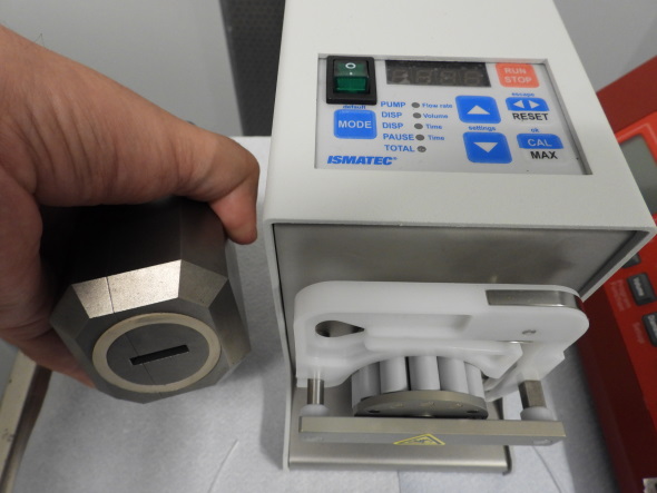
Swisstrace twilite blood sampling system
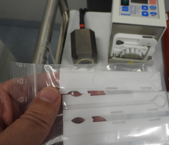
Swisstrace twilite is one of the commercially available automatic blood sampling systems (ABSS) that enables measurement of arterial input function (AIF) with temporal resolution of 1 second. In small animal studies it can be used to avoid blood loss due to surgically placed arteriovenous shunt. It is MR-compatible, and can also be used to assess AIF in human subjects (Zhang et al., 2014). This device, with a femoral arteriovenous shunt, has provided AIF with better accuracy than image-derived input function in mice (Alf et al., 2013a) and in rats (MacAskill et al., 2019).
To obtain accurate analysis results of the PET data, the blood cell uptake of the radioligand needs to be accounted for (Alf et al., 2013b; Bolcaen et al., 2016). Dispersion in the sampling system can be measured and corrected as described by Convert et al (2007). Sufficient shunt flow rate in required to minimize the dispersion error (Warnock et al., 2011).
When Swisstrace twilite ABSS is operated via PMOD PSAMPLE, the software corrects the data for the background, branching ratio and decay of the isotope, and calibrates the data into units kBq/cc. The system clocks of the twilite acquisition computer and PET scanner should be synchronized for accurate decay correction.
The calibrated and decay corrected blood curve (BTAC) can be saved as .crv file,
which is a tab-delimited text file which can be easily imported in Excel or used by TPC tools.
If needed, it can be converted to some other file formats using
tacformat.
Dispersion correction, conversion of the blood curve to plasma and correction for metabolites can be done in PMOD PKIN tool, or using the command-line programs and scripts from TPC.
See also:
Literature
Alf MF, Wyss MT, Buck A, Weber B, Schibli R, Krämer SD. Quantification of brain glucose metabolism by 18F-FDG PET with real-time arterial and image-derived input function in mice. J Nucl Med. 2013a; 54(1): 132-138. doi: 10.2967/jnumed.112.107474.
Alf MF, Martić-Kehl MI, Schibli R, Krämer SD. FDG kinetic modeling in small rodent brain PET: optimization of data acquisition and analysis. EJNMMI Res. 2013; 3: 61. doi: 10.1186/2191-219X-3-61.
Bolcaen J, Lybaert K, Moerman L, Descamps B, Deblaere K, Boterberg T, Kalala JP, Van den Broecke C, De Vos F, Vanhove C, Goethals I. Kinetic modeling and graphical analysis of 18F-Fluoromethylcholine (FCho), 18F-Fluoroethyltyrosine (FET) and 18F-Fluorodeoxyglucose (FDG) PET for the discrimination between high-grade glioma and radiation necrosis in rats. PLoS One 2016; 11(8): e0161845. doi: 10.1371/journal.pone.0161845, correction: 10.1371/journal.pone.0164208.
Rey-Bretal D, Moscoso A, Gómez-Lado N, Fernández-Ferreiro A, Silva-Rodríguez J, Ruibal Á, Aguiar P. Feasibility of longitudinal brain PET with real-time arterial input function in rats. Mol Imaging Biol 2021; 23: 350-360. 10.1007/s11307-020-01556-y.
Warnock G, Bahri MA, Goblet D, Giacomelli F, Lemaire C, Aerts J, Seret A, Langlois X, Luxen A, Plenevaux A. Use of a beta microprobe system to measure arterial input function in PET via an arteriovenous shunt in rats. EJNMMI Res. 2011; 1(1): 13. doi: 10.1186/2191-219X-1-13.
Updated at: 2021-12-24
Created at: 2019-12-03
Written by: Vesa Oikonen